By virtue of my specialization in pediatric ophthalmology, strabismus and neuro-ophthalmology, I get to see a large number of children with mental retardation and visual impairment In my clinic. In a lot of such situations, this is their first visit to an ophthalmologist as the parents have been busy with managing the developmental delay and have been visiting the paediatrician. It is generally when a paediatrician or paediatric neurologist sees them and refers them to me that the parents begin to realise there is something wrong with the child’s eyes. In a contrast to this presentation, there are cases where the parents notice that the child does not seem to be making eye contact with them or seems disinterested in his/her surroundings or has an obvious eye abnormality such as squint (misaligned eyes) or cataract (white reflex in the eyes), and bring the child to me first. It is on examination that I realise there is a generalised developmental delay and the problem encompasses a lot more than an eye disease alone. These cases are then referred to the paediatric neurologist who evaluates the child in detail. Either way, both cases require a comprehensive multi-disciplinary management strategy and are a challenge for rehabilitation therapists and physicians. In this article, I will briefly touch upon the ophthalmologists’ perspective to visual rehabilitation of a child with mental retardation through the help of a case I had recently encountered in my clinic.
A 14 month old boy was referred to me by my paediatric neurologist colleague with history of hypoxia at birth resulting in generalised developmental delay. The child was born to a non-consanguineous marriage and had a relatively uneventful antenatal life in the mother’s womb. The mother was a young primipara in her early twenties and lived in Gwalior, India. She was taken up for a induced vaginal delivery at a nursing home in her town as she had reached full term. Unfortunately during the induction, a prolonged labour ensued and there was fetal distress with meconium stained liquor. Since the nursing home did not have adequate facility for an emergency caesarean section at the time, the delivery was completed with assistance of forceps. The child did not cry at birth and was noted to be cyanosed though there was no written record documenting this. The history I took from the child’s family clearly pointed towards a limp, lifeless baby who needed to be resuscitated and began to cry after a few minutes of birth. After the delivery, everything seemed uneventful and the child was feeding well though less interactive. It was around 6 months of age that the family started to note that the child was “slow” and could not hold the neck or fixate at any object. The eyes seemed to dance around but they thought this would get better and their paediatrician asked them to wait for some more time. By the time the child was 1 year old, he could hold his head but could barely sit with support but not without it and could vocalise with sounds but no words. The child’s eyes would still dance around and the child could not fixate at either the mother’s face or any object. Some jerky head movements were also noted but seizures were ruled out. The child was then seen by a pediatric neurologist who advised an MRI and found gliotic changes in the brain and diagnosed it as a case of generalised development delay secondary to hypoxic ischemic encephalopathy. The child was also sent for an ENT exam where the BERA test was normal (indicating a normal hearing). The child was also sent to me for an ophthalmic evaluation.
I examined the child when the child was fully awake and alert and found that the child could perceive light from my torch but could not fixate or follow it. There was a nystagmus (wriggly dancing eye movements) in both eyes. I tried testing the vision of the child with the use of special charts called Lea gratings but the child seemed to be unable to fixate at them. I understood that the child has a poor vision and I had to understand the reason for it. The next step in examine the child was to evaluate the pupillary reflexes in both the eyes and they seemed to be brisk and normal. This indicated to me that there was unlikely to be a problem with one of the eyes’ nerves (optic nerves). After this, I dilated the child’s pupils with an eyedrop to look for presence of cataract and any pathology in the retina or nerve. I found all these to be normal. I understood that the cause for vision loss was likely related to the brain problem (encephalopathy). One last thing that we checked before completing the examination was the child’s refraction (or power of the eye) by retinoscopy. To my surprise, I found that the child had a very high plus power, in the range of +10 in both eyes. This made me think that the child may possibly have an ametropic amblyopia (lazy eye). However, I knew at this stage that a normal eye of a 1 year old child can also be a power of upto +5 or+6 and that I have to repeat the retinoscopy examination under atropine cycloplgia to get the true power of this child. I then explained to the parents that the child needs another checkup after putting eye ointment atropine and that we may have to prescribe glasses. The parents were very worried and reluctant to the idea of giving glasses to such a small child. I made them understand that if the child does not wear glasses when the power is so high, the eyesight will be very poor and the eyes will develop amblyopia (become a lazy eye) which will be difficult to treat at a later age. I also made them realise that if the child sees better and nystagmus reduces, then the child will be more likely to interact with his environment and this will greatly benefit the mental development. After this, the parents agreed and came back to me after 1 week after having put atropine in the child’s eyes. This time when we performed the retinsocopy test, we found that the power of the child’s eyes was +12 Dioptre and I prescribed glasses of the same number. The parents had lots of questions such as how will we ensure that the child wears the glasses, what type of glasses to use, how will the glasses stay on the child’s face, what if the child breaks the glass etc. I answered each of these questions for them. I told them that it was their duty to make the child wear the glasses and as long as the child is wearing the glasses, there will be improvement in vision and for every few hours that the child will not be wearing glasses, there will be a delay in improvement. I told them that children’s spectacles are special and are made of light, relatively unbreakable materials and have a band to tie them at the back of the head. I asked them to get the lenses made of polycarbonate material so that the lenses do not break even if the child was to throw the glasses around or something was to hit the child’s face. Finally I advised them to try the glasses with an open mind and see the difference it will make to their child. Convinced but still a bit reluctant, the parents accepted my advice and made a pair of glasses for the child.
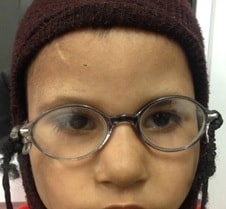
The child came back to me after 1 month of using the glasses. The parents told me that the child had initially been irritated and tried to remove the glasses the moment they were place on the face but with the parents’ perseverance and occasional force and distraction, the child had started using the glasses in everyday. The parents told me that they were surprised to see the child adaprt so quickly and so well to the glasses that now if they removed the glasses for any reason, the child cried. I emphasised to them that the spectacles were giving the child a better vision and so the child liked wearing them. This is in fact a lesson to be learnt: if the proper power of glasses is given, most children will adapt to them very well and actually better than adults.
We let the child continue to use the glasses regularly. Over the next 6 months, the nystagmus seemed to reduce and the child could fixate better and started to interact better with his surroundings. There was a steadier gaze and as the child was more and more stimulated by the environment, there was improvement in mental status too. This was an excellent example of how the correction of 1 sensory system led to overall improvement in development.
At the last follow up, the child was 4 years old, able to stand, walk and speak basic sentences. The power of glasses had reduced by 1 Dioptre and nystagmus was significantly better.
In conclusion, this case demonstrates the need to evaluate all sensory systems when dealing with a child with developmental delay. It also highlights the need to optimise the sensory inputs to achieve better overall development. On the ocular rehabilitation front, it highlights the need for proper refraction, early prescription of glasses and need to counsel parents to ensure acceptability of spectacles.
![DigvijayProfile[1]](https://drdigvijaysingh.com/wp-content/uploads/2017/11/DigvijayProfile1.jpg)